
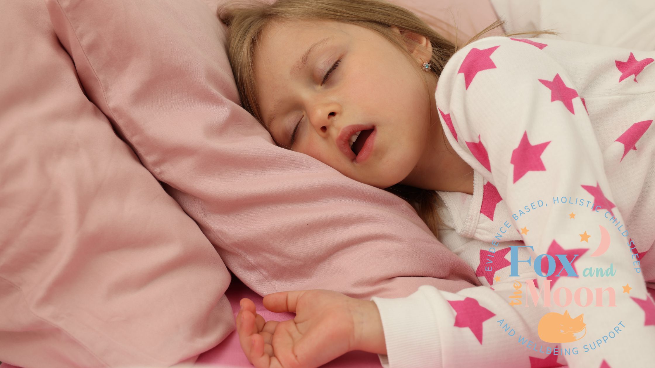
Joining the Dots Nobody Joined: Infant Feeding, Oral Function and the Long Road to the ENT Waiting Room
By Emily Whalley — A hypothesis piece for practitioners and curious parents
What if some of the most common childhood health problems we treat, recurrent ear infections, enlarged tonsils and adenoids, sleep-disordered breathing, even some presentations of ADHD, have roots in the first hours of a baby’s life? Not in genetics, not in bad luck, but in something as specific as whether anyone properly assessed how that baby feeds?
This is not a claim that the evidence has already proven. It is something more interesting than that: a hypothesis, built from several independent bodies of research that have, until now, remained in separate silos. This piece is an attempt to join those dots, carefully, honestly, and with full acknowledgement of where the evidence is strong, where it is suggestive, and where the research simply has not yet been done.
It begins, perhaps surprisingly, in a French maternity ward.
What Other Countries Are Already Doing Differently
France has long integrated osteopathy into its maternity care in a way that has no equivalent in the United Kingdom. In many French hospitals, osteopathic sessions are offered during the maternity stay itself, organised either in a dedicated space within the ward, or directly in the mother’s room (Hôpital Franco-Britannique, 2017). This is not fringe practice. It reflects a cultural and clinical understanding that birth, however uncomplicated, places significant mechanical forces on the newborn body, forces that may affect the cranium, the cervical spine, the jaw, and the soft tissues of the oral cavity.
Brazil takes a different but equally instructive approach. In 2014, Brazil became the first country in the world to enshrine neonatal oral function assessment in law. Under Law 13.002, all newborns in every public and private hospital and maternity ward must undergo a lingual frenulum assessment, the so-called *Teste da Linguinha*, or tongue test, before discharge (Martinelli, Marchesan & Berretin-Felix, 2014). This assessment, developed by speech-language pathologist Roberta Martinelli, evaluates both the anatomy and the function of the lingual frenulum within the first 48 hours of life. It sits alongside four other mandatory neonatal screenings: pulse oximetry for congenital heart disease, red reflex for eye disorders, hearing screening, and the Guthrie test for metabolic diseases (PLOS One, 2021). Oral function, in Brazil’s national health policy, is considered a core public health priority on a par with cardiac and sensory screening.
Norway and Switzerland, meanwhile, consistently record among the lowest rates of gastro-oesophageal reflux disease of any country globally. According to Global Burden of Disease 2021 data, Switzerland has the lowest GERD incidence of any nation studied (age-standardised rate of 2,091 per 100,000), and Norway has the lowest prevalence (4,330 per 100,000), compared with countries like Brazil, El Salvador and the Dominican Republic which sit at the extreme high end of the burden (PMC, 2025).
These facts, taken in isolation, are interesting. Taken together, they begin to suggest something worth examining more carefully.
The Clinical Chain: From Feeding Difficulty to Downstream Harm
To understand what may be connecting these international patterns, it is necessary to trace a clinical pathway that runs from the earliest days of life through to outcomes that most clinicians would not instinctively connect to infant feeding.
### Step One: Oral Function and the Unassessed Newborn
Ankyloglossia, tongue tie, occurs in approximately 3.2% to 4.8% of consecutive term infants at birth, rising to 12.8% in infants presenting with breastfeeding difficulties (Academy of Breastfeeding Medicine, 2004). These figures vary considerably depending on the assessment tool used and the training of the assessor, which is itself part of the problem: there is no universally accepted diagnostic criterion, and identification is inconsistent even within a single healthcare system.
What is less contested is that tongue tie, when present and functionally significant, disrupts the mechanics of feeding. Effective sucking requires the tongue to lift, cup, and generate negative intraoral pressure. When the lingual frenulum restricts these movements, the infant compensates, and compensation has consequences. One proposed mechanism, described by Siegel (2016) in a retrospective analysis of 1,000 breastfeeding dyads, is aerophagia: the excessive swallowing of air that results from a dysfunctional latch. This aerophagia-induced reflux presents clinically in ways that are indistinguishable from gastro-oesophageal reflux disease, and in Siegel’s analysis, was being treated with acid-suppressing medication that addressed the symptom while leaving the cause entirely untouched.
Crucially, the oral examination that might identify this cause is not routinely performed in UK neonates. Current guidance from most expert bodies recommends assessment only when breastfeeding problems are already presenting, a reactive, rather than preventive, model. Brazil’s mandatory approach represents the alternative: assess every baby before the problem has time to compound.
Step Two: The Reflux That May Not Be Reflux
The medicalisation of infant reflux has accelerated dramatically over the past two decades. In France, 6.1% of children under two years were using proton pump inhibitors in 2019, up from 3.6% in 2010 (Taine et al., 2021). Comparable increases have been documented across Europe and beyond. Yet the evidence base for PPI use in infants is strikingly weak: multiple randomised controlled trials have found no difference between PPIs and placebo for crying and irritability in infants with reflux (van der Pol et al., 2011). This raises an important question: if the medication is not working, is the diagnosis correct?
When aerophagia — driven by dysfunctional oral mechanics, is the primary driver of symptoms, acid suppression is not merely ineffective. It may be actively harmful. Studies have found that PPI use in infants is associated with increased rates of upper respiratory infections, gastroenteritis, and community-acquired pneumonia (Canani et al., 2006). The altered gastric pH creates an environment more permissive to pathogenic colonisation, and the underlying functional problem, the tongue, the latch, the suck, remains entirely unaddressed.
There is also a second, underappreciated mechanism at work. Research by Vanderbilt University found that infants with reported GERD had significantly higher bronchiolitis severity scores than those without, nearly twice the odds of clinically meaningful disease severity after adjustment for confounders (HEI et al., 2014). Whether this association reflects aspiration of gastric contents, shared inflammatory pathways, or some other mechanism remains unclear. But the clinical implication is significant: a baby who is refluxing, for whatever reason, is a baby who is more vulnerable when respiratory viruses arrive.
Step Three: Respiration, the Upper Airway, and the ENT Pathway
Recurrent otitis media, adenoidal hypertrophy, and chronic upper airway inflammation are well-recognised sequelae of persistent reflux in infants and young children. Acid and pepsin in the upper airway provoke an inflammatory response that contributes to Eustachian tube dysfunction, middle ear effusion, and the hypertrophy of lymphoid tissue, the tonsils and adenoids, that the body uses as its first line of immunological defence.
This matters because tonsillectomy and adenoidectomy (T&A) rates vary enormously between countries in ways that are not explained by underlying disease burden alone. In 1998, paediatric T&A rates ranged from 19 per 10,000 children in Canada to 118 per 10,000 in Northern Ireland (van den Akker et al., 2004). A 2016 analysis of OECD data across 31 countries found that healthcare system type was a significant driver of variation, but even controlling for system type, the variation persisted, suggesting that underlying disease burden is genuinely different between populations (Bhattacharyya & Kepnes, 2016).
What nobody has yet examined is whether countries with better neonatal oral function identification and feeding support have lower rates of childhood upper airway surgery. This is the gap in the literature that this piece is proposing exists, and that it is worth closing.
Step Four: Sleep, Neurodevelopment, and the ADHD Question
Adenotonsillar hypertrophy is the leading cause of obstructive sleep apnoea in children. Sleep-disordered breathing in early childhood, even at subclinical levels that do not meet formal apnoea criteria, is associated with significant neurodevelopmental consequences, including impairments in attention, executive function, and behavioural regulation (Bhattacharjee et al., 2012).
These are, of course, the defining features of ADHD.
The association between breastfeeding duration and ADHD risk is now supported by multiple independent datasets. A South Korean nationwide study of over 1.1 million infants found that exclusive breastfeeding was associated with a 23% reduction in the odds of subsequent ADHD diagnosis, compared with exclusive formula feeding, with a dose-response pattern suggesting a genuine biological relationship rather than confounding alone (ADHD Evidence, 2023). An Israeli retrospective study found that children bottle-fed at three months were three times more likely to develop ADHD than those who were breastfed at the same age (Mimouni-Bloch et al., 2013). A US nationally representative dataset found that breastfeeding for 6–12 months was associated with 57% lower prevalence odds of ADHD diagnosis, after controlling for twelve potential confounders (Soled et al., 2021).
The mechanism through which breastfeeding exerts this protective effect is not yet established. It may be the nutritional composition of breast milk, long-chain polyunsaturated fatty acids supporting myelination, iron bioavailability supporting dopaminergic function. It may be the act itself, the sustained oral-motor work of breastfeeding supporting craniofacial development and airway patency. It may be both. What is clear is that breastfeeding duration is shorter in families where oral function problems are unidentified and unsupported, and that ADHD rates are higher in children who were not breastfed.
Whether tongue tie → feeding difficulty → shortened breastfeeding → increased ADHD risk is a genuine causal chain, or whether the association between breastfeeding and ADHD is explained by other shared factors, remains unknown. But the hypothesis is coherent, the individual links are evidenced, and it has not been studied as a connected pathway.
Why the Dots Have Not Been Joined
The silos that separate these bodies of evidence are partly disciplinary and partly structural. Neonatologists do not typically track their patients’ ENT outcomes at age six. Paediatric ENT surgeons do not typically take neonatal feeding histories. ADHD clinicians rarely ask about breastfeeding duration, let alone lingual frenulum assessment at birth. And the researchers studying each of these questions are publishing in entirely different journals, attended by entirely different professional communities.
There is also a deeper issue. The outcomes being discussed here, T&A rates, ADHD diagnoses, respiratory admission rates, are measured at the country or healthcare system level, and are influenced by so many upstream variables that isolating the effect of neonatal oral function identification would require a study of considerable complexity and duration. Breastfeeding rates correlate with socioeconomic status, maternal education, cultural norms, and workplace policy, all of which independently affect the outcomes in question. T&A rates are driven heavily by surgeon propensity and healthcare system type, as much as by underlying pathology. ADHD diagnosis rates vary internationally by diagnostic criteria and cultural attitudes towards the diagnosis, not just by true prevalence.
These confounders are real and should be named honestly. They do not, however, invalidate the hypothesis. They define the shape of the study that would be needed to test it.
What a Connected Approach Might Look Like
Countries like France and Brazil offer natural experiments. France has embedded osteopathic newborn assessment into its maternity culture, addressing birth-related cranial and oral strain early, supporting feeding mechanics from the outset. Brazil has mandated neonatal lingual frenulum screening for every infant born in the country. Neither country has, to date, tracked whether these interventions are associated with downstream reductions in childhood ENT surgery, respiratory admissions, or neurodevelopmental diagnoses.
But they could. And the data, in many cases, already exists, it is simply not being connected.
A joined-up research programme might ask: in populations where neonatal oral function is routinely assessed and supported, whether through mandatory screening, osteopathic maternity care, lactation consultant integration, or some combination, do we see differences in:
– Duration of exclusive breastfeeding
– Rates of infant reflux diagnosis and PPI prescribing
– Bronchiolitis severity and hospital admission rates
– Rates of recurrent otitis media and middle ear surgery
– Paediatric T&A rates
– ADHD diagnosis rates at school age
Each of these outcomes is measurable. The countries in which to measure them already exist. The gap is the will, and the interdisciplinary framing, to look across them together.
The ADHD Pathway Is Already Pointing Backwards — Nobody Has Noticed Yet
Perhaps the most striking piece of evidence that this hypothesis has clinical legs comes not from neonatology or ENT, but from the ADHD assessment pathway itself, and specifically from a change quietly embedded in American paediatric guidelines.
The American Academy of Pediatrics’ 2019 Clinical Practice Guideline for ADHD, the most widely referenced framework for ADHD assessment in children, includes a key action statement requiring clinicians to screen for physical comorbidities as part of every ADHD evaluation. Explicitly listed among those physical conditions is sleep apnoea (American Academy of Pediatrics, 2019). This is not a minor footnote. It is a formal clinical requirement, carrying a Grade B strength of recommendation, that sleep-disordered breathing must be considered before an ADHD diagnosis is confirmed and treatment initiated.
The evidence underpinning this requirement is substantial. A meta-analysis of studies examining the relationship between sleep-disordered breathing and ADHD in paediatric populations found that the two conditions are significantly associated, and, critically, that adenotonsillectomy was associated with a meaningful reduction in ADHD symptom scores, with a medium effect size across twelve studies (Sedky et al., 2014). The conclusion of that meta-analysis was unambiguous: treatment of comorbid sleep-disordered breathing should be considered before medicating ADHD symptoms if sleep-disordered breathing is present.
The scale of potential misdiagnosis here is not trivial. The incidence of sleep-disordered breathing among children with ADHD is estimated at between 25% and 57% (Sedky et al., 2014). One study found that 65% of children with ADHD had a diagnosable sleep disorder, compared with just 17% of controls (Joseph et al., 2022, cited in PMC, 2025). Research in mouth-breathing children, a population with significant overlap with those who have unresolved upper airway dysfunction, found they may be 40 to 100 times more likely to develop behavioural problems resembling ADHD than nasal-breathing peers, leading researchers to explicitly recommend screening for mouth breathing before any ADHD diagnosis is made (PMC, 2021).
Children who are sleep-deprived do not present as tired. They present as hyperactive, impulsive, and inattentive, because an exhausted child’s nervous system responds to insufficient restorative sleep with dysregulation, not drowsiness. This is not widely understood outside specialist settings, and it means that a child whose upper airway has been quietly obstructed for years may accumulate a constellation of symptoms that maps precisely onto the diagnostic criteria for ADHD, and may be medicated accordingly, with stimulants that can further disrupt sleep, while the structural cause of their presentation remains entirely unaddressed.
What is not in any current guideline, not in the AAP’s ADHD pathway, not in any ENT or sleep medicine framework, is any instruction to take a neonatal feeding history. No guideline currently asks: was this child’s oral function assessed at birth? Was there a tongue tie? Were there breastfeeding difficulties that went unsupported? How long was this child breastfed, and why did breastfeeding end?
These questions are not asked because the pathway connecting them to the child now sitting in an ADHD assessment clinic has never been formally described. This is that description.
The Pathway, Complete
Stated plainly, the hypothesis this piece is proposing runs as follows:
A baby is born with a functionally significant lingual frenulum restriction that is not identified, because no routine oral function assessment is performed. Breastfeeding is difficult from the start. The baby compensates with a dysfunctional suck pattern, swallowing excess air. Symptoms of discomfort, frequent waking, and apparent pain are attributed to reflux. A PPI is prescribed. The oral cause remains unaddressed.
Shortened by difficulty and pain, breastfeeding ends earlier than the family had hoped. The baby misses months of the oral-motor exercise, craniofacial stimulation, and nutritional composition that breastfeeding provides. The upper airway, less well-developed than it might have been, is more susceptible to the chronic inflammatory processes that persistent reflux promotes. Eustachian tube dysfunction follows. Adenoidal hypertrophy follows. The child has recurrent ear infections, then grommets, then T&A surgery that improves their sleep but does not fully resolve the neurodevelopmental consequences of years of fragmented, unrestorative rest.
At seven years old, they are referred for ADHD assessment.
The AAP guideline now requires that a sleep study is considered before medication is initiated. That sleep study may well identify residual sleep-disordered breathing. Treatment of that breathing may improve symptoms significantly. But the opportunity to interrupt this pathway at its very beginning, in a maternity ward, in the first 48 hours of life, with a simple, non-invasive, low-cost oral function assessment, has long passed.
This is not a counsel of despair. Children can be helped at every point along this pathway. But the earlier the intervention, the less compound the harm. And the earliest possible point is the one that currently receives the least systematic attention.
A Note on What This Is and Is Not
This piece is a hypothesis, not a systematic review. The individual evidence strands cited here are real and peer-reviewed, but the chain that connects them has not been formally studied as a unified pathway. The argument being made is that it should be, not that it has been proven.
Practitioners reading this are encouraged to hold it as a framework for clinical thinking rather than a protocol for practice. The implication for individual clinical encounters is modest and uncontroversial: assess oral function early, support feeding proactively, and consider the downstream effects of unresolved feeding difficulty as extending well beyond the immediate neonatal period.
The implication for research is more ambitious: that we are missing something significant by failing to study the first hours of a baby’s life as the beginning of a pathway that extends, for some children, all the way to the ENT theatre, the sleep clinic, or the ADHD assessment room.
The American Academy of Pediatrics has already, perhaps without fully realising it, pointed the arrow in the right direction. The ADHD assessment pathway now looks backwards towards sleep. Sleep looks backwards towards the upper airway. The upper airway looks backwards towards infant feeding. Infant feeding looks backwards towards oral function at birth.
Someone just needs to follow it all the way.
References
Academy of Breastfeeding Medicine (2004). *ABM Clinical Protocol #11: Guidelines for the Evaluation and Management of Neonatal Ankyloglossia and its Complications in the Breastfeeding Dyad*. Breastfeeding Medicine.
American Academy of Pediatrics (2019). Clinical Practice Guideline for the Diagnosis, Evaluation, and Treatment of Attention-Deficit/Hyperactivity Disorder in Children and Adolescents. *Pediatrics*, 144(4), e20192528.
Bhattacharyya, N. & Kepnes, L.J. (2016). Variation in tonsillectomy rates by health care system type. *International Journal of Pediatric Otorhinolaryngology*, 88, 163–167.
Bhattacharjee, R. et al. (2012). Adenotonsillectomy outcomes in treatment of obstructive sleep apnea in children. *American Journal of Respiratory and Critical Care Medicine*, 185(9), 994–1002.
Canani, R.B. et al. (2006). Therapy with gastric acidity inhibitors increases the risk of acute gastroenteritis and community-acquired pneumonia in children. *Pediatrics*, 117(5), e817–e820.
Global Burden of Disease Collaborative Network (2025). Socioeconomic perspective on the global burden of gastroesophageal reflux disease (1990–2021). *PMC / Frontiers in Public Health*.
Hôpital Franco-Britannique (2017). *Osteopathy at the Maternity Ward*. Available at: hopitalfrancobritannique.org.
Martinelli, R.L.C., Marchesan, I.Q. & Berretin-Felix, G. (2014). Lingual frenulum protocol with scores for infants. *International Journal of Orofacial Myology*, 38, 104–112.
Mimouni-Bloch, A. et al. (2013). Breastfeeding may protect from developing attention-deficit/hyperactivity disorder. *Breastfeeding Medicine*, 8(4), 363–367.
PMC (2021). Sleep difficulties and symptoms of attention-deficit hyperactivity disorder in children with mouth breathing. *PMC*, 8645617.
PMC (2025). Polysomnographic insights into the attention-deficit/hyperactivity disorder and obstructive sleep apnea connection in children. *Frontiers in Sleep*.
PLOS One (2021). Prevalence of the five newborn screening tests. *PLOS One*, 16(9).
Sedky, K., Bennett, D.S. & Carvalho, K.S. (2014). Attention deficit hyperactivity disorder and sleep disordered breathing in pediatric populations: a meta-analysis. *Sleep Medicine Reviews*, 18(4), 349–356.
Siegel, S.A. (2016). Aerophagia induced reflux in breastfeeding infants with ankyloglossia and shortened maxillary labial frenula (tongue and lip tie). *International Journal of Clinical Pediatrics*, 5(1), 6–8.
Soled, D. et al. (2021). Breastfeeding is associated with a reduced risk of attention-deficit/hyperactivity disorder among preschool children. *Journal of Developmental and Behavioral Pediatrics*, 42(1), 9–15.
Taine, M. et al. (2021). Paediatric outpatient prescriptions in France between 2010 and 2019. *Lancet Regional Health – Europe*, 7, 100129.
van den Akker, E.H. et al. (2004). Large international differences in (adeno)tonsillectomy rates. *Clinical Otolaryngology*, 29(2), 161–164.
van der Pol, R.J. et al. (2011). Efficacy of proton-pump inhibitors in children with gastroesophageal reflux disease: a systematic review. *Pediatrics*, 127(5), 925–935.
Emily is the founder of Fox & The Moon Sleep. This piece represents a hypothesis-in-progress, not clinical guidance. The author welcomes correspondence from researchers and clinicians working in related fields